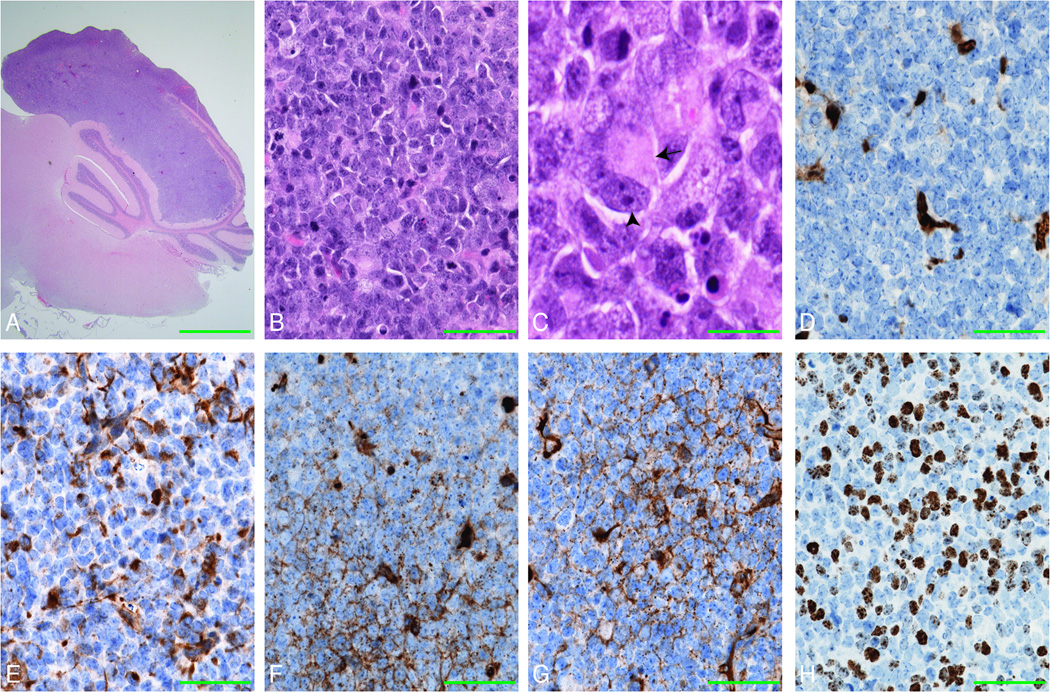
Figure 6. Snf5F/Fp53L/LGFAP-Cre mice develop CNS AT/RT.
(A–C) H&E stained sagittal section of a 5 months old mouse Snf5F/Fp53L/LGFAP-Cre brain, demonstrates a tumor infiltrating the cerebellum with obliteration of the folia (A). The tumor high grade, poorly differentiated tumor (B) characterized by the presence of cells with eccentrically placed nuclei, prominent nucleoli (arrowhead) and eosinophilic inclusion-like cytoplasm (arrow), resembling rhabdoid cells (C) in a background of non-descriptive primitive neuroectodermal cells.
(D–H) Ini1/Baf47 expression (D) is loss in the nuclei of all tumor cells, but is retained in the vascular endothelial elements (brown). Focal expression (brown) of cytokeratin (E), synaptophysin (F), and GFAP (G) and high nuclear expression of Ki67 (H) was also observed in the tumor cells. Scale bars represent 800 µm (A), 40 µm (B, D-H) and 27 µm (C).

