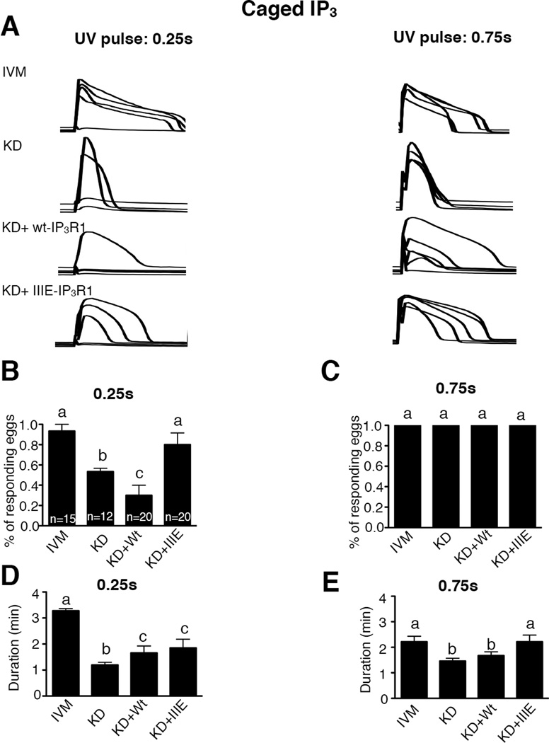
Figure 5. CDK- and ERK-related phosphorylations on IP3R1 enhance the sensitivity of IP3R1 in mouse oocytes.
(A) Changes in [Ca2+]i induced by photolysis of cIP3 in IVM, KD, KD+wt-IP3R1 and KD+IIIE-IP3R1 mouse eggs. Two UV pulses with different duration (0.25s and 0.75s) were conducted sequentially and noted in each of the panels. The number of responding eggs expressed in % (B,C) and the duration of the Ca2+ release (D,E) caused by a 0.25s and 0.75s UV pulse were compared among the different treatments mentioned above. Bars with different superscripts represent treatments that are significantly different (P<0.05).

