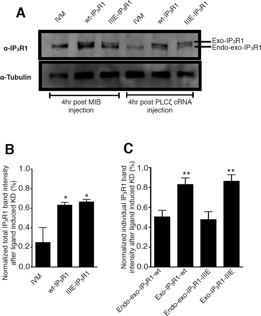
Figure 6. Differential PLCζ-induced degradation rate of endogenous and exogenous IP3R1s in mouse oocytes.
(A) Immunoblotting of egg lysates from IVM eggs, wt-IP3R1 and IIIE-IP3R1 expressing eggs 4hr after MIB or PLCζ cRNA injection, probed with the Rbt03 antibody and anti–α-tubulin antibody. (B) Intensity of the total IP3R1 signal including the upper (Exo-IP3R1) and lower bands (Endo-exo-IP3R1) from panel A 4 hr after PLCζ cRNA injection was calculated. (C) Intensity of the Exo-IP3R1 and Endo-exo-IP3R1 bands was calculated separately in the groups of 6A. In both panels B and C, the amount present in IVM, wt-IP3R1 and IIIE-IP3R1 eggs after injection with MIB was chosen as control and set at 100%; the band intensities observed in IVM, wt-IP3R1 and IIIE-IP3R1 eggs injected with PLCζ cRNA were presented relative to the control condition.

