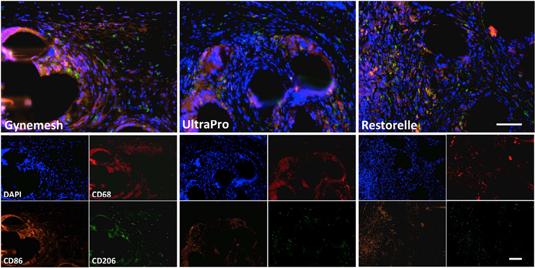
Figure 3.
Immunofluorescent labeling with antibodies to CD68 (Pan-macrophage, red), CD86 (M1 macrophage, orange), CD206 (M2 macrophage, green) and DAPI (nuclei, blue) is shown. Few positive cells were observed in sham-operated animals (not shown). Predominance of the M1 macrophage response was observed in Gynemesh PS, UltraPro and Restorelle groups. Combined fluorescent channels are shown in the top panel, and individual channels in the bottom panel. All images at 20× magnification, scale bars = 100 µm.

