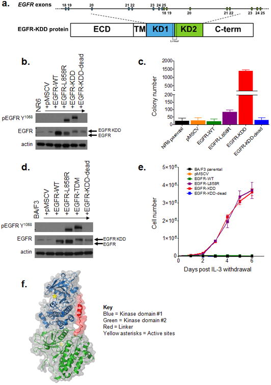
Figure 1. The EGFR-KDD is an oncogenic EGFR alteration.

(a) Schematic representation of EGFR-KDD depicting the genetic and protein domain structures. ECD = extracellular domain. TM = transmembrane domain. Blue = EGFR exons 18-25 #1. Green = EGFR exons 18-25 #2. KD1 = first kinase domain. KD2 = second kinase domain. C-term = carboxyl terminus. (b) Representative western blot of NR6 cells stably expressing indicated EGFR constructs. EGFR-KDD-dead is a kinase dead version of EGFR-KDD. (c) NR6 cells stably expressing the indicated constructs (pMSCV = vector only) were plated in triplicate in soft agar, grown for 15 days, and quantified for colony formation. (d) Representative western blot of BA/F3 cells expressing indicated EGFR constructs. (e) BA/F3 cells transfected with indicated constructs (pMSCV = vector only) were grown in the absence of IL-3 and counted every 24 hours. (f) Ribbon diagram and space-filling model of the EGFR-KDD kinase domains (GLY 696 - PRO 1370) illustrating the proposed mechanism of auto-activation. Blue = first kinase domain; green = second kinase domain; red = linker; yellow asterisks = active sites.
