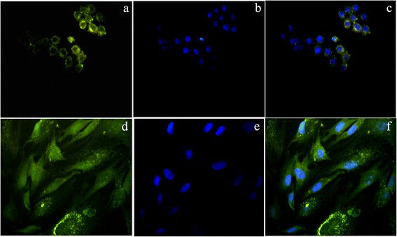
Figure 6.

Cellular imaging-fluorescence microscopy: (a & d) HeLa and RADMSC cells showed the efficient cellular internalization of QDs in the cytoplasm and nearby nucleus [yellowish green fluorescene]; (b & e) Nucleus were counterstained blue with DAPI [blue flurescene]; (c & f) Overlay of QDs and DAPI fluorescene images.
