
Fig. 3.
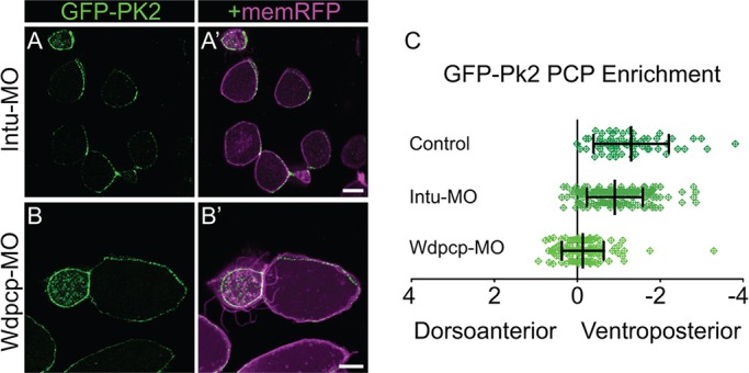
Wdpcp knockdown disrupts core PCP patterning. (A,B) Mosaically labeled epidermal cells in St.31 X. laevis embryos have GFP-Pk2 localized asymmetrically upon Intu-MO knockdown (A) and symmetrically upon Wdpcp-MO knockdown (B). (C) Quantification of PCP enrichment shows a significant shift upon Intu-MO knockdown (P=0.0031, n=187 cells) and a more significant shift upon Wdpcp-MO knockdown (P<0.0001, n=137 cells) in comparison to controls (n=64 cells). Error bars indicate s.e.m. Scale bars: 10 μm.