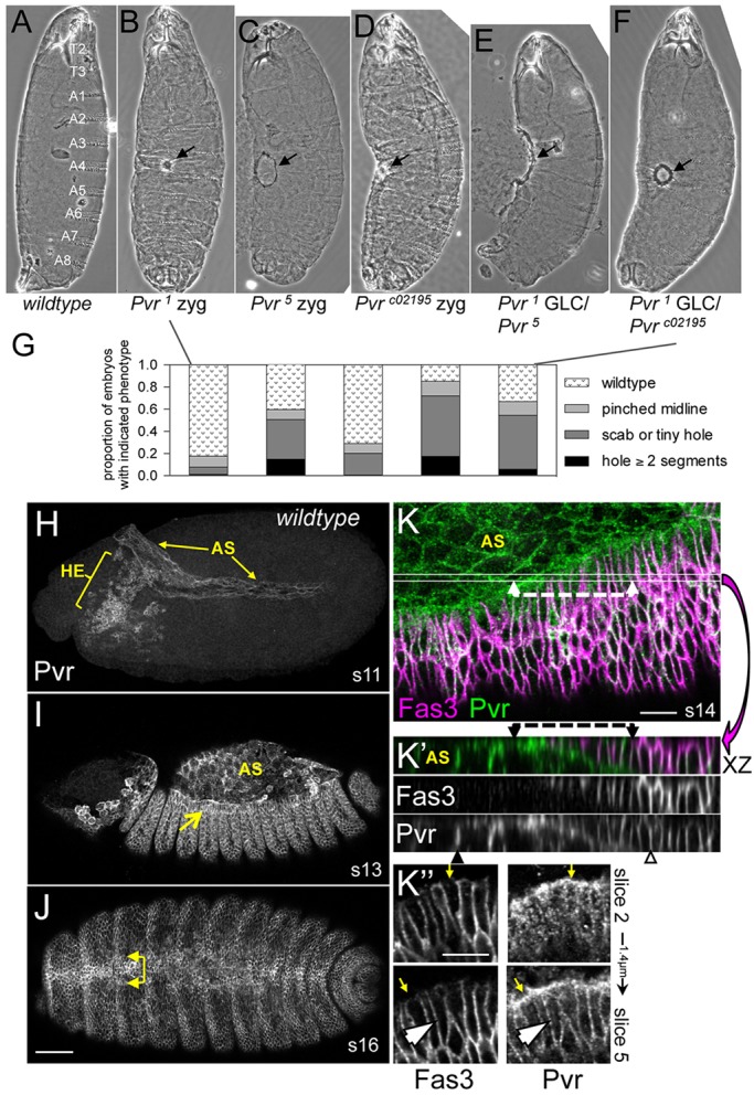
Fig. 1.

Embryonic closure phenotypes and protein expression of Pvr. (A-F) Larval cuticles. Wild type, with the segments indicated (A), is compared with Pvr zygotic (B-D) or germline clone (GLC) (E,F) mutants displaying midline scabs or holes (arrows). (G) Quantification of phenotypes in B-F (n=124-310). (H-K″) Pvr immunostaining in wild-type embryos, without (H-J) or with (K-K″) Fas3 colocalization. Germband extended (H), early dorsal closure (I) and post-closure (J) embryos depict Pvr expression in hemocytes (HE), amnioserosa (AS) and leading edge (LE) of the dorsal ectoderm (arrows, I,J). (K) Confocal z-projection of dorsal quadrant from mid-closure embryo. Thin lines demarcate the position of the x/z projection beneath (K′); single channels are also shown. Dashed lines highlight Pvr localization at the interface between LE and underlying AS. Note Pvr at lateral membranes of AS (black arrowhead). Ectoderm has extensive colocalization of markers (open arrowhead). Planar polarized enrichment of Pvr is seen in LE cells compared with Fas3 (K″, arrows). Lateral membrane colocalization is seen in the deeper section (arrowhead). Scale bars: 50 μm in H-J; 10 μm in K-K″.
