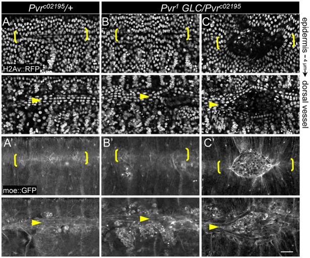
Fig. 3.
Cardia bifida in Pvr mutants. (A-C) Single confocal sections of epidermis or dorsal vessel showing the indicated fluorescent markers in live embryos. Late-stage heterozygotes (A,A′) have closed (midline bracketed) and cardiac cells have coalesced underneath in an aligned double row (arrowheads). Pvr mutants have mild (dorsal vessel only, B,B′) or moderate (C,C′) ectodermal hole and split vessel phenotypes. Scale bar: 20 μm.

