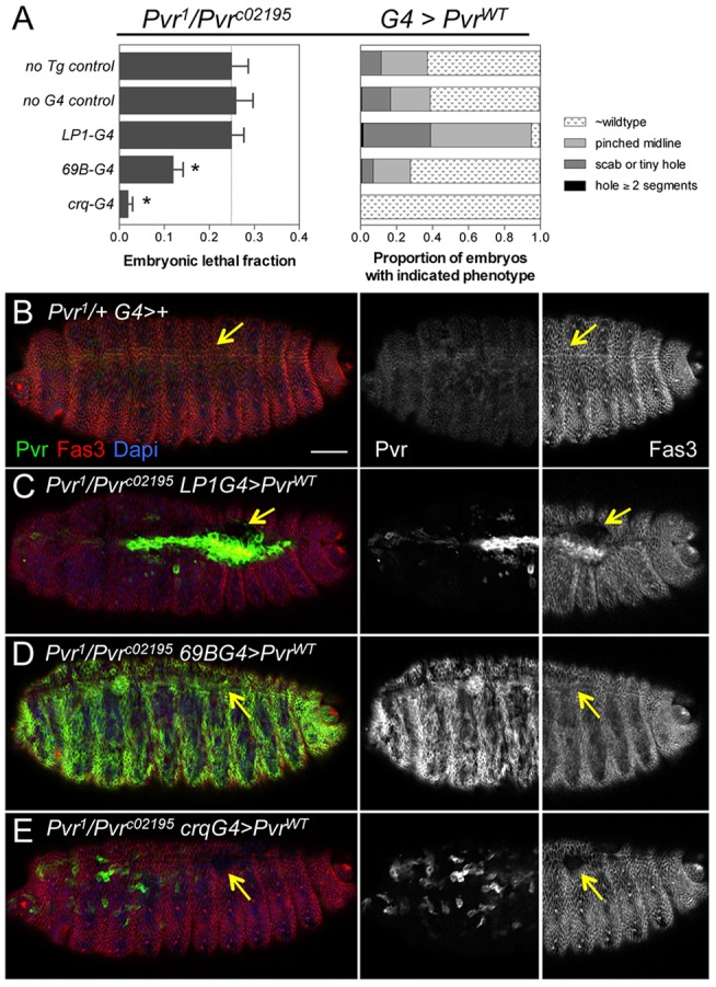
Fig. 4.

Complementation of Pvr in embryonic hemocytes rescues lethality. (A) Quantification of embryonic lethal fraction (left) and cuticle phenotypes (right) from tissue-specific rescue crosses. The lethal fraction was significantly reduced by expression of Pvr in ectodermal derivatives (69B-G4) or hemocytes (crq-G4). Bars show mean±s.d. *P<0.0001 (Chi-square test). n=269-394. Line indicates the expected fraction (0.25) for homozygous recessive embryonic lethal mutation. (B-E) Pvr immunostaining detects endogenous and overexpressed protein in merged and single-channel images (embryo anterior). Fas3 is shown in merged and righthand panels (embryo posterior). Arrows point to the dorsal midline, which is closed in similarly staged control (B) and 69B-G4 (D) embryos, but open in LP1-G4 (C) and crq-G4 (E) embryos. Scale bar: 20 μm.
