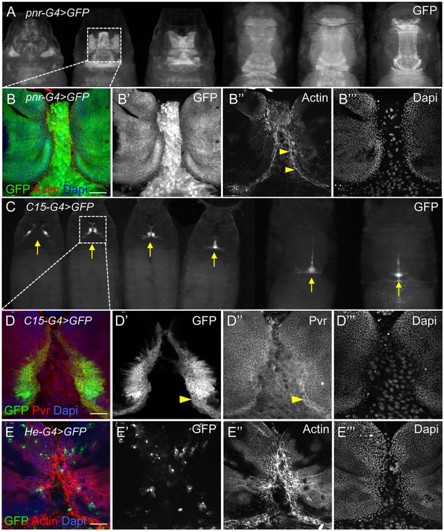
Fig. 9.
Pvr expression at the LE of spreading imaginal discs during thorax closure. (A,C) GFP expression with pnr-G4 (A) or C15-G4 (C) drivers in progressively older live pupae reveals apposition and fusion of notum tissue at the midline (arrows) during thorax closure. Boxes indicate regions shown as dissected, fixed tissues in panels beneath. (B-B‴) pnr drives expression in discs (small packed nuclei) in a broad proximal domain and in the intervening larval epidermal tissue (large nuclei). DAPI staining shows nuclei (blue). F-actin is enriched at the LE margins (arrowheads). (D-D‴) GFP expression in the C15 domain is restricted to a narrow region of the disc margins. Endogenous Pvr protein is enriched in mesenchymal-like marginal cells (arrowheads). (E-E‴) For comparison, the He-G4 driver directs GFP expression in hemocytes infiltrating the tissues. Actin protrusions are prominent, knitting together disc margins. Scale bars: 50 μm.

