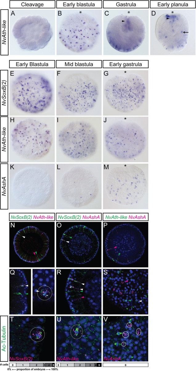
Fig. 2.
NvSoxB(2), NvAshA and NvAth-like are co-expressed during development, but NvAshA is not found in mitotic cells. (A-D) Expression of NvAth-like is first detected in early blastulae, in scattered cells that are absent from one region of the embryo (presumptive endodermal plate) (B). In gastrulae (C), expression is in scattered ectodermal cells predominantly in the aboral region of embryos, also in the developing pharynx (arrowhead). These domains are similar in early planulae (D), with the addition of newly expressing cells in the endoderm (arrow). Whereas NvSoxB(2) (E-G) and NvAth-like (H-J) are already expressed in early blastula stages, NvAshA (K-M) is expressed in only a few cells by mid-blastula stage, and is not broadly expressed until the early gastrula. (A,C,D) Mediolateral optical sections; (B,E-M) surface views. (N-V) Double FISH at blastula and gastrula stages shows some co-localisation of NvSoxB(2) with both NvAth-like and NvAshA (N,O,Q,R), but minimal co-localisation between NvAth-like and NvAshA (P,S). Based on the elongated shape of the DAPI nuclear staining, mitotic cells expressing both NvSoxB(2) and NvAth-like were identified (dashed circle in Q). By staining spindles with an acetylated tubulin antibody (green), we observed mitotic cells expressing NvSoxB(2) (T) and NvAth-like (U) in single FISH (dashed circles). NvAshA transcripts (circled in V) were never found in cells undergoing mitosis. Charts show how many mitotic, gene-expressing cells were observed in each of 20 early- and mid-gastrula embryos/gene. White arrowheads, co-localisation; green and pink arrowheads, single transcript localisation. Blue, DAPI; Ac-Tubulin, anti-acetylated tubulin. Asterisk, oral pole.

