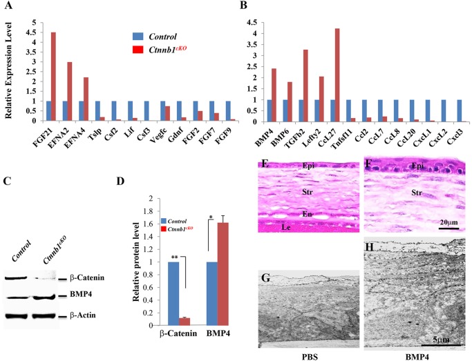
Fig. 5.
Differential expression of growth factors/cytokines upon Ctnnb1 ablation. (A,B) Ad-Cre-GFP-treated or Ad-GFP-treated primary cell cultures of stromal keratocytes isolated from Ctnnb1f/f mice at P0 were screened using a mouse cytokine primer library. The Ctnnb1cKO mutant showed changes in the expression of various cytokines and growth factors, including a 2.5-fold increase in Bmp4. (C) Western blotting analyses confirmed the increase in Bmp4 after Ctnnb1 ablation. β-actin provided a loading control. (D) Statistical analysis of western blot results showed that Bmp4 expression was increased at the protein level. (E-H) Histological analysis of corneas from P10 mice treated with recombinant BMP4 (10 ng/ml) or PBS (control) every other day from P0 to P8. H&E staining showed that Bmp4 administration results in an increase in the number of epithelial cells (3-4 cell layers; compare E with F). Likewise, TEM showed that BMP4 administration resulted in a much thicker epithelium (H) than that of the control (G). *P<0.05, **P<0.01. Mean±s.e.m.

