Fig. 1.
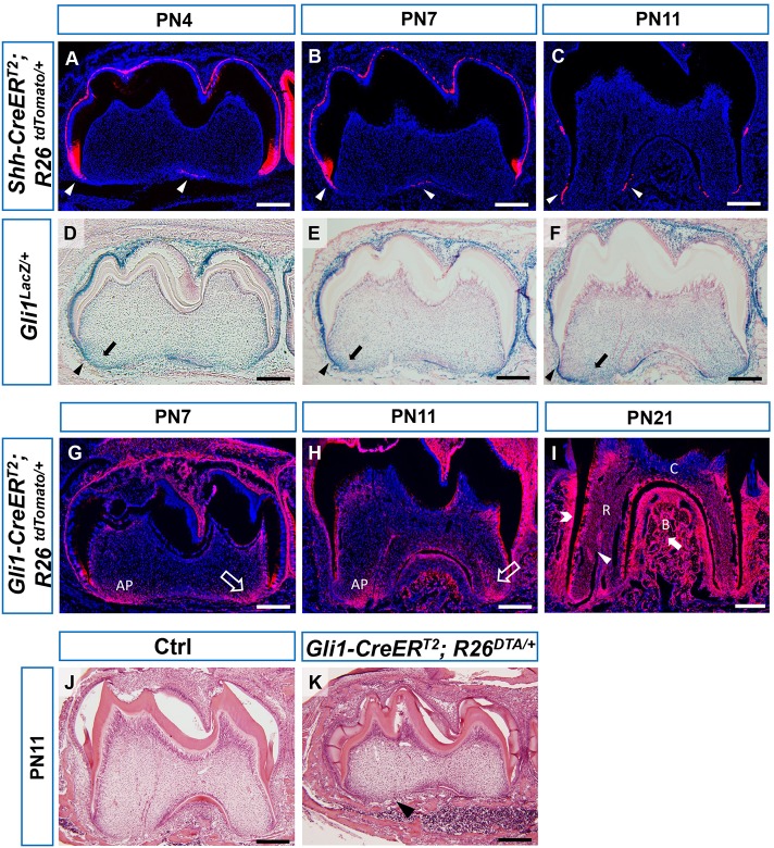
Gli1+ cells are root progenitor cells. (A-C) Sagittal sections of first mandibular molars of PN4, PN7 and PN11 Shh-CreERT2;R26tdTomato/+ reporter mice 30 h after tamoxifen injection. Arrowheads indicate fluorescent signal in the dental epithelium. (D-F) X-gal staining of first mandibular molars of PN4, PN7 and PN11 Gli1lacZ/+ mice. Arrowheads indicate strong signal in dental epithelium. Arrows indicate signal in apical papilla. (G-I) Lineage tracing of Gli1+ cells in first mandibular molars of PN7, PN11 and PN21 Gli1-CreERT2;R26tdTomato/+ mice after tamoxifen injection at PN3. Arrows indicate labeling of apical papilla in G-H. Arrowhead, open arrowhead and solid arrow indicate labeling of root pulp, PDL and alveolar bone, respectively. (J,K) HE staining of PN11 control (Ctrl) and Gli1-CreERT2;R26DTA/+ mice treated with tamoxifen at PN3. Arrowhead indicates lack of root formation. n=3 per group. AP, apical papilla; C, crown; R, root; B, alveolar bone. Scale bars: 200 µm.

