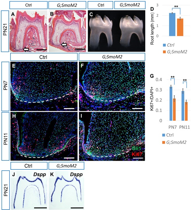
Fig. 3.
Constitutive activation of Hh signaling in root progenitor cells leads to reduced proliferation and shorter roots. (A-C) HE staining and macroscopic views of first mandibular molars from PN21 control (Ctrl) and Gli1-CreERT2;R26SmoM2fl/fl (G;SmoM2) mice after tamoxifen injection at PN3. Arrows indicate the apical end of the mesial root. (D) Quantification of the sum of the mesial and distal root lengths of first mandibular molars at PN21 from micro-CT. n=3 per group. (E,F,H,I) Immunostaining of Gli1 (green) and Ki67 (red) in the mesial apical papilla of first mandibular molars from PN7 and PN11 control and G;SmoM2 mice. Dotted lines indicate the HERS, dashed lines indicate the border of the apical papilla. (G) Quantification of Ki67+ nuclei/total nuclei from E,F and H,I. n=3 per group. (J,K) In situ hybridization of Dspp in the first mandibular molars of PN21 control and G;SmoM2 mice. Scale bars: 500 µm in A,B,J,K; 100 µm in E,F,H,I. **P<0.01.

