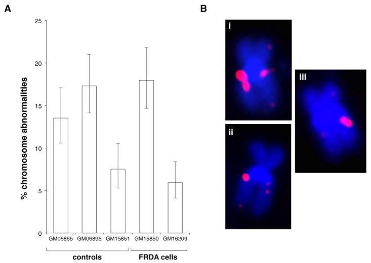
Fig. 1. Both normal and FRDA cells show a high frequency of spontaneous chromosomal abnormalities in the region of the FXN gene.
A) The percentage of abnormal chromosomes seen in normal and patient lymphoblastoid cells. B) Examples of the chromosomal abnormalities observed in these cells. (i) Chromosome with a duplication on one sister chromatid. (ii) Chromosome with a deletion on one sister chromatid. (iii) Chromosome with a duplication on one chromatid and a deletion on the other. Chromosomes were stained with DAPI (blue) and FXN signal is shown in red. Chromosomal duplications made up 76–90% of the chromosomal abnormalities with the remainder being deletions of some or all of the probe region.

