Fig. 2.
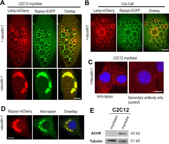
Rapsyn concentrates at junctional sites between vacuolin-1 enlarged lysosomal vacuoles. (A) Undifferentiated C2C12 myoblasts or (B) non-muscle COS-7 cells were transfected with rapsyn–EGFP and Lamp1–mCherry and either left untreated (lower panels) or treated with vacuolin-1 (upper panels). Rapsyn–EGFP colocalizes perfectly with Lamp1–mCherry in the juxtanuclear region of non-treated C2C12 myoblasts (A, lower panels) and is concentrated at the junctional sites between enlarged lysosomal vacuoles visualized by Lamp1–mCherry (A, upper panels) and in COS cells (B). (C) C2C12 myoblasts were treated with vacuolin-1, fixed and labeled with mouse monoclonal anti-rapsyn antibody followed by a secondary fluorescent antibody (left panel) or with a fluorescent secondary antibody only (right panel). (D) C2C12 myoblast transfected with rapsyn–mCherry, fixed and labeled with antibody against rapsyn. Scale bars: 10 µm. (E) Blot showing that AChRs are undetectable in undifferentiated myoblasts.

