Fig. 4.

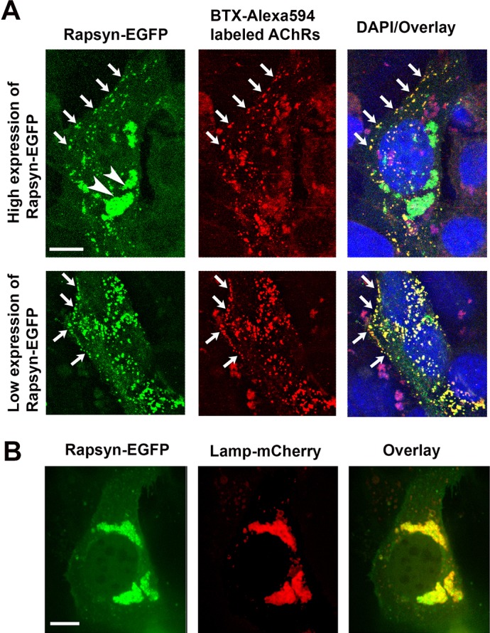
Rapsyn is recruited to the cell surface in the presence of AChRs. C2C12 myoblasts were co-transfected with rapsyn–EGFP and either (A) AChRα, AChRβ, AChRδ and AChRε subunits or (B) Lamp1–mCherry. (A) In C2C12 myoblasts expressing AChRs and high levels of rapsyn–EGFP, rapsyn is concentrated in the juxtanuclear region (arrowheads) and colocalizes with AChR clusters at the cell surface (upper panel). However, in myoblast expressing AChRs and low level of rapsyn, most rapsyn molecules are recruited to AChR clusters at the cell surface (lower panel). Arrows indicate co-clusters of AChRs and rapsyn at the cell surface. (B) When myoblasts were transfected with rapsyn–EGFP and Lamp1–mCherry, rapsyn remains largely concentrated with Lamp1–mCherry in the juxtanuclear region (no visible accumulation at the cell surface). Scale bars: 10 µm.
