Fig. 8.
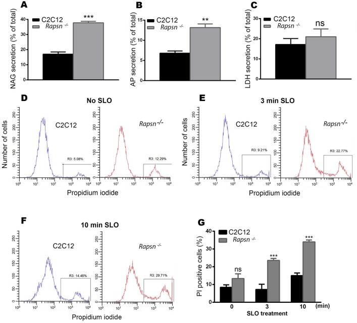
Lysosomal exocytosis was significantly increased in Rapsn−/− myoblasts. Graphs showing lysosomal enzyme activities. (A) β-N-acetylglucoseaminidase (NAG), (B) acid phosphatase (AP) and (C) Lactate dehydrogenase (LDH). Note that both lysosomal enzymes were significantly increased in rapsyn-deficient myoblasts compared to C2C12 myoblasts. Results are mean±s.e.m. (D–F) Representative histograms obtained by FACS analysis of propidium iodide staining of live cells: (D) non-treated C2C12 myoblasts (left) and Rapsn−/− (right) myoblasts; (E) treated (permeabilized) myoblasts exposed to 500 µg/ml of bacterial toxin streptolysin-O (SLO) for 3 min or (F) 10 min. In each histogram, the left peak corresponds to intact (propidium iodide negative) cells and the right peak corresponds to propidium-iodide-positive cells. Note that untreated Rapsn−/− myoblasts show a higher basal uptake of propidium iodide than C2C12 cells, indicative of plasma membrane damage. This difference in propidium iodide uptake becomes larger as cells are challenged with SLO for 3 min (E) or 10 min (F), indicating that in the absence of rapsyn, plasma membrane is more vulnerable to damage. (G) Quantification of FACS data (as shown in D,E, and F), which is reported as percentage of propidium-iodide-positive Rapsn−/− and C2C12 myoblasts that were either untreated (0 min) or treated with SLO for the indicated periods of time. The data represent mean±s.e.m. from three independent experiments.

