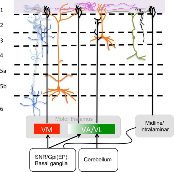
Figure 2.

Motor thalamus (MT) and related midline/intralaminar thalamic connections to layer 1. Depicted in the rectangles at the bottom are projections from MT divided according to a direct VM and rostromedial VA projection to L1 and a caudolateral VA/VL projection that does not reach L1 as described by Kuramoto et al. (2015) (see “MT Inputs” Section ) illustrated also is the minor input from midline/intralaminar nuclei to L1 via the basal ganglia (see “Midline/Intralaminar Nuclei” Section). Specific L1 neurons are depicted neurogliaform (purple) and single bouquet (black) (see “Layer 1: The Crowning Enigma” Section). The rectangle at the top marks where axons from specific neurotransmitter producing nuclei (i.e., ACh, NE, 5HT and DA) run along L1. Sketched are also important neuronal processes mingled in L1 from axons of Martinotti cells (blue), vertical dendrites from bipolar interneurons that run horizontally once in layer 1 (green) and apical dendritic tufts of layers 2/3 and 5 pyramidal cells (orange) (see “Neuronal Processes that Mingle in Layer 1” Section). For accurate afferent arborizations consult Arbuthnott et al. (1990), Kuramoto et al. (2009) and Cruikshank et al. (2012).
