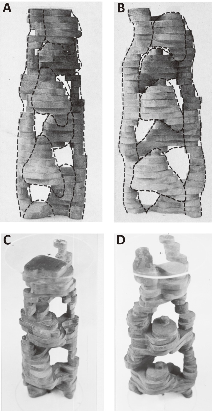
Figure 2.
Solid models of actin-containing filament reconstructed from electron micrographs of negatively stained filaments by assuming the helical symmetry of specimens.34) A. A front view of a solid model of the complex of actin, tropomyosin, troponin-T, and troponin-I. It represents an inhibited state of thin filaments. B. A front view of actin-tropomyosin representing an active state of thin filament. C. An oblique view of the model shown in A (inhibited state). D. An oblique view of the model shown in B (active state). In an inhibited state, actin and tropomyosin associate more firmly.

