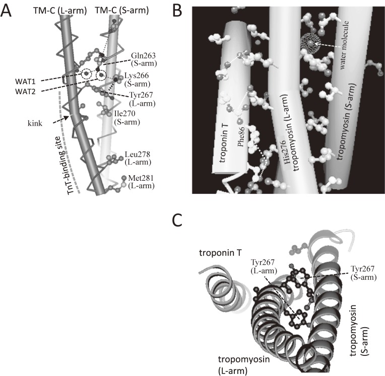
Figure 13.
Asymmetry of the tropomyosin coiled coil as determined by X-ray crystallograpy.119) A. In a coiled-coil structure of COOH-region of tropomyosin in the binary complex, one of α-helix (L-arm) was kinked. One water molecule in the hydrophobic core facilitated asymmetry and labeled as WAT1, whereas the other water molecule is labeled as WAT2. B. Troponin-T binds to the kinked region of L-arm. The water molecule in the hydrophobic core was also observed in the ternary complex. The planar side chain of His-276 of TM-C (L-arm) face against that of Phe-86 (cardiac Phe-110) of TnT. C. The top view from the M-line side. Two Tyr-267s of L-arm and S-arm near the hydrophobic core responsible for the kink formation are shown in ball-and-stick format.

