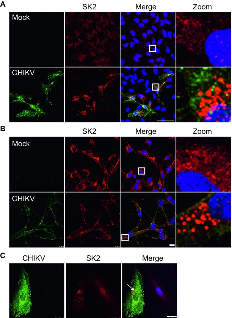
Figure 3.
SK2 is re-localized during CHIKV infection. HeLa (A), hSKMC (B), or MCF10A (C) cells were uninfected or infected with CHIKV at a MOI of 5 for 24 h. The viral E2 protein (green), SK2 (red), and Hoechst nuclear stain (blue) were visualized by confocal microscopy. Scale bar = 50 µm (A), 10 µm (B), 25 µm (C).

