
FIGURE 1.
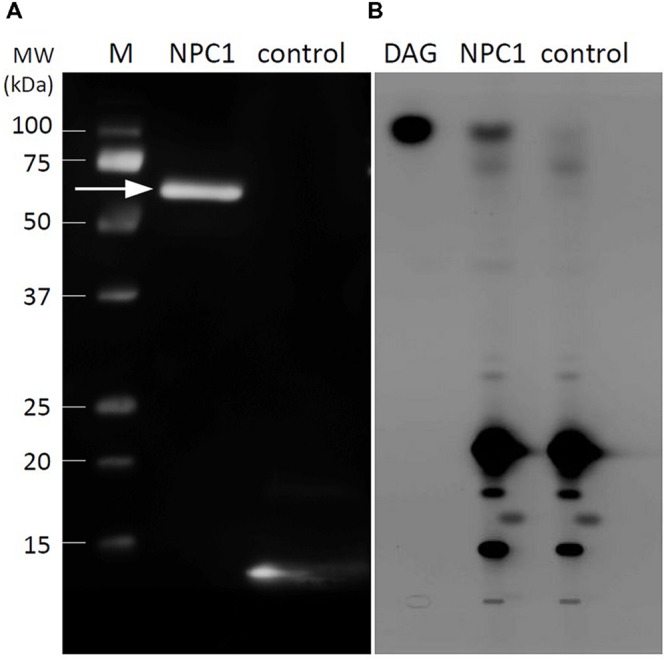
Western blot analysis and in vitro activity of recombinant NPC1. (A) Immunoblotting analysis of recombinant NPC1 protein after purification, desalting and concentration. The arrow indicates the 64 kDa protein corresponding to NPC1. The empty vector pET30 served as a control. Part of the expression region of the empty vector is transcribed with 6xHis and detected as a band of small size. (B) Activity assay of recombinant NPC1 protein and vector-only control performed with bodipy-PC as a substrate. Twenty micrograms of protein per assay was used. After the extraction of lipids and separation by high-performance thin layer chromatography, DAG was observed in NPC1. M, marker; DAG, diacylglycerol; NPC1, non-specific phospholipase C1.