Figure 2.

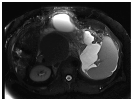
Selected magnetic resonance imaging frame showing a large peri-pancreatic pseudocyst extending from the pancreatic tail to the anterior abdominal wall in a patient with pancreatitis and splenic vein thrombosis.

Selected magnetic resonance imaging frame showing a large peri-pancreatic pseudocyst extending from the pancreatic tail to the anterior abdominal wall in a patient with pancreatitis and splenic vein thrombosis.