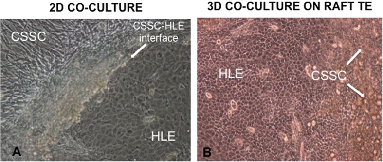
Figure 1.

(a) Light microscopy image showing a primary culture of a mixed population of human corneal stromal stem cells (CSSC) and human limbal epithelial cells (HLE) on plastic (passage 0, day 15). Spontaneous organization of the two different cell types is observed (CSSC-HLE interface). The characteristic cobblestone morphology of a HLE cluster is visible adjacent to confluent CSSC at its periphery (magnification ×10). (b) Light microscopy image showing a mixed population of human corneal stromal stem cells (CSSC) and human limbal epithelial cells (HLE) cultured on RAFT TE for 13 days. A confluent monolayer of epithelial cells with the characteristic cobblestone morphology is visible with clumps of CSSC (white arrows) shedding from the surface of RAFT TE (magnification ×10).
