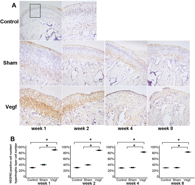
Figure 6. Expression of vascular endothelial growth factor receptor 2 (VEGFR2) in the condyle cartilage in the control, sham, and vegf groups at weeks 1, 2, 4, and 8.
(A) Histological analysis of VEGFR2-positive chondrocytes. VEGFR2-positive chondrocytes are distributed in all cartilage layers in the vegf group from week 1 onwards. (B) Comparison of the percentage of VEGFR2-positive chondrocytes in the hypertrophic layer between the groups. The percentage of VEGFR2-positive chondrocytes is significantly higher in the vegf group than in the control and sham groups at all time points (*P < 0.05).

