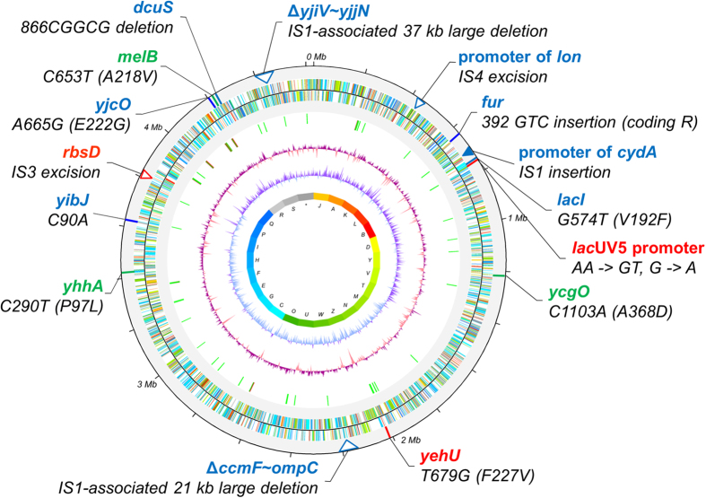
Figure 1. Genomic variations in E. coli C41(DE3) and C43(DE3) as compared to the BL21(DE3) backbone sequence in a circular representation.
Mutations found in both C41(DE3) and C43(DE3) are depicted in red, C41(DE3) in green, and C43(DE3) in blue, respectively. Small-scale changes like SNPs or small DIPs are indicated with solid lines, and IS element-mediated large scale insertions or deletions with triangles.

