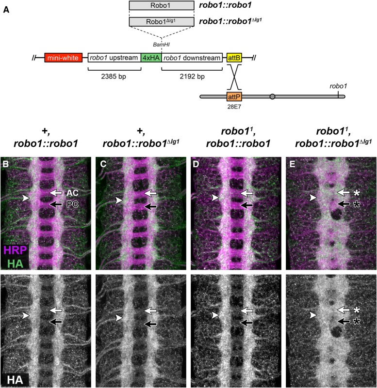
Figure 4.
Expression of Robo1 and Robo1ΔIg1 proteins via a robo1 genomic rescue transgene. (A) Schematic of robo1 rescue construct. Open reading frames are cloned into the BamHI restriction site in-frame with the N-terminal 4xHA epitope tag and are expressed under the control of robo1 genomic regulatory sequences. Rescue constructs carrying full-length Robo1 or Robo1ΔIg1 coding sequences were inserted into the same genomic landing site at cytological position 28E7. robo1 mutations were introduced onto these chromosomes via meiotic recombination. (B–E) Stage 16 embryos stained with anti-HRP (magenta) and anti-HA (green) antibodies. Bottom images show HA channel alone from the same embryos. (B, C) In a wild-type background, HA-tagged full-length Robo1 (B) or Robo1ΔIg1 (C) proteins expressed from the robo1 rescue transgene are localized to longitudinal axon pathways (arrowhead) and are excluded from commissural axon segments in both the anterior commissure (AC, white arrow) and posterior commissure (PC, black arrow). The HA staining pattern in both embryos closely matches the expression of endogenous Robo1 protein in wild-type embryos (compare to Figure 3E). (D) Embryos homozygous for a null allele of robo1 and carrying two copies of the robo1::robo1 rescue construct display a wild-type axon scaffold, and the distribution of HA-tagged Robo1 is the same as in a wild-type background. (E) Homozygous robo1 mutants carrying two copies of the robo1::robo1ΔIg1 transgene exhibit a robo1 loss of function phenotype, with thickened commissures and longitudinal pathways that form closer to the midline. HA-tagged Robo1ΔIg1 is detectable on longitudinal pathways (arrowhead) and also on both commissures (arrows with asterisks), although Robo1ΔIg1 levels appear higher on AC (white arrow with asterisk) than PC (black arrow with asterisk).

