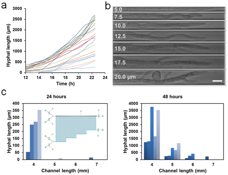
Figure 3. Hyphal growth and morphology on the microfluidic chips.
(a) Growth curves of 22 individual hyphae growing in the channels (2.5 mm × 10 μm × 10 μm) on a single chip. The hyphal length is measured at regular intervals of 30 min starting 12 h after conidial trapping. (b) Images of hyphae growing in 7 different widths (5, 7.5, 10, 12.5, 15, 17.5 and 20 μm) of channels (2.5 mm long). Scale bar, 10 μm. (c) Hyphal extension in 4 different lengths (4, 5, 6 and 7 mm) of channels (10 μm wide) with 5 replicates for each length after 24 hours (left) and 48 hours (right). Inset shows a schematic of chip design. In the experiments, N. Crassa strain NMF617 expressing histone H1-RFP is grown under the constant flow of 10% glucose in 1× Vogels’ salts solution at 0.3 μL/min.

