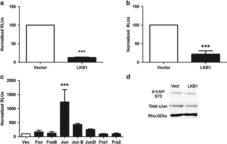
Figure 5.
LKB1 expression is associated with decreased MMP-1 and AP-1 activity. (a) MDA-MB-231 cells were transiently transfected with LKB1 or vector control and MMP-1-luciferase plasmid for 24 h. Cells were lysed and luciferase levels measured. Bars represent relative light units (RLUs) normalized to vector control cells±s.e.m. of triplicate experiments. (b) MDA-MB-231-vector or MDA-MB-231-LKB1 cells were transfected with AP-1-luciferase plasmid for 24 h. Cells were lysed and luciferase levels measured. Bars represent RLUs normalized to vector control cells±s.e.m. of triplicate experiments. (c) MDA-MB-231-LKB1 cells were transiently transfected with expression plasmids for each individual AP-1 family member (Fos, FosB, Jun, JunB, JunD, Fra1, Fra2) or vector control and MMP-1-luciferase plasmid for 24 h. Cells were lysed and luciferase levels measured. Bars represent RLUs normalized to vector control cells±s.e.m. of triplicate experiments. ***P<0.001. (d) Western blot analysis of MDA-MB-231-vector or -LKB1 stable cells for phospho-(S73) and total c-Jun. Rho-GDIα serves as a loading control. Blot representative of three independent experiments.

