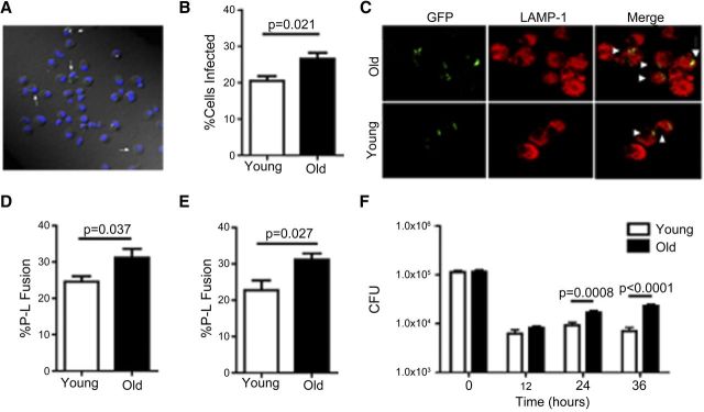
Figure 2. Pulmonary macrophages from old mice have increased M.tb uptake and P-L fusion but a defect in intracellular control of bacterial growth.

Pulmonary macrophages isolated from old (solid bars) and young (open bars) mice were adhered to coverslips and incubated with 5:1 GFP-M.tb for 2 h and analyzed by confocal microscopy (A–E). White arrows indicate infected pulmonary macrophages (A). Percentage of cells from both groups found to be infected with GFP-M.tb (B). P-L fusion was observed in macrophages from young and old mice using the lysosomal marker LAMP-1 (C and D) and cathepsin D (E). White arrowhead indicates P-L colocalization events (yellow) between GFP-M.tb (green) and LAMP-1 (red). Macrophages isolated from the lungs of young and old mice were incubated with 5:1 GFP-M.tb for 2 h, and M.tb intracellular growth was assessed at the indicated time-points postinfection (F). CFU was normalized between macrophages from young and old mice at time zero. Colonies were enumerated after 21 days of incubation at 37°C. Data were combined from three independent experiments from pools of five mice. Student's t-test was used to determine statistical significance.
