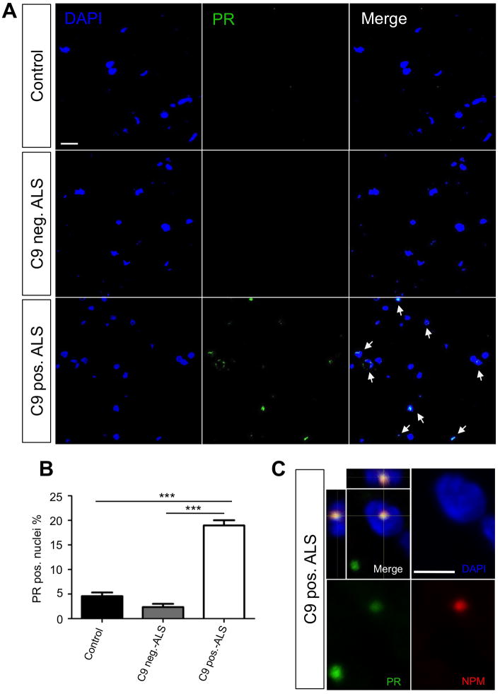
Figure 8. Nuclear PR inclusions are found in C9ORF72 ALS/FTD patient spinal cord tissues.
(A) Representative images of a 15 slice z-series performed on age-matched human lumbar spinal cord slices from various patient types (control, C9ORF72 mutation negative ALS, and C9ORF72 mutation positive ALS). PR dipeptides (green), DAPI (blue), and merged images show absent to low intensity staining in control and C9ORF72 mutation negative ALS tissues. C9ORF72 mutation positive tissues shows higher intensity staining as well as nuclear localization of the PR dipeptides. (B) Quantification of PR positive nuclei was performed on 20 15-slice z-series images for each patient type with ~400 nuclei quantified. C9ORF72 mutation positive tissues showed a significantly higher (P< 0.0001) percent of PR positive nuclei (18.96 ± 1.07) than control (4.464 ± 0.88) and C9ORF72 mutation negative ALS (2.44 ± 0.67). (C) Immunofluorescence analysis shows that PR aggregates co-localize with nucleophosmin (NPM) in the nucleus from C9ORF72 mutation positive patient spinal cord tissues. DAPI: blue; PR: green; NPM: red. Calibration bar is 5 μm.

