Figure 5.
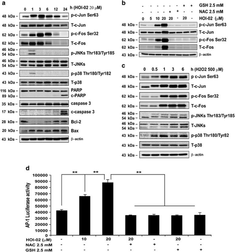
HOI-02-induced apoptosis is mediated through induction of AP-1 activity and the modulation of caspase expression. (a) KYSE510 cells (1 × 10⁶) were treated with 20 μM HOI-02 for the indicated times. The expression of apoptosis-related proteins was determined by western blot analysis using specific antibodies. β-Actin was detected in the same membrane and served as a loading control. Data shown are representative of results from triplicate independent experiments. (b) KYSE510 cells (1 × 10⁶) were treated with various doses of HOI-02 (0, 5, 10 or 20 μM), 2.5 mM NAC or 2.5 mM GSH for 24 h. The expression of proteins was determined by western blot analysis using specific antibodies. (c) ROS induce phosphorylation of c-Jun and c-Fos in esophageal cancer cells. After treatment with 500 μM H2O2 for the indicated times, the expression of proteins was determined by western blot analysis using specific antibodies. (d) HOI-02 induces AP-1 luciferase activity and the effect is blocked by NAC or GSH in KYSE510 esophageal cancer cells. KYSE510 cells were transiently transfected with the AP-1 luciferase reporter gene construct and incubated with HOI-02 (0, 10 or 20 μM), 2.5 mM NAC or 2.5 mM GSH. Luciferase activity was measured as described in Materials and methods section

