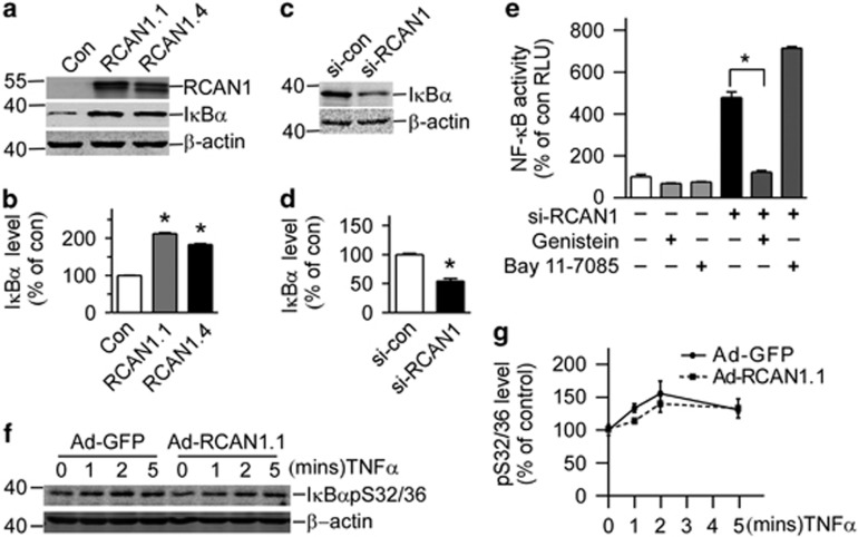
Figure 2.
RCAN1 increased the endogenous IκBα levels (a) RCAN1 increases the level of IκBα. HEK293 cells were transfected with pcDNA3.1RCAN1.1/1.4-6myc. Endogenous IκBα levels were detected by anti-IκBα antibody. RCAN1 protein was detected by anti-myc antibody. β-actin detected by anti-β-actin was used as loading controls. (b) Quantification of a. Values represent mean±S.E.M.; n=3; *P<0.0001 by Student's t-test. (c) HEK293 cells were transfected with si-RCAN1; endogenous IκBα levels were detected by anti-IκBα antibody. (d) Quantification of c. Values represent mean±S.E.M.; n=3; *P<0.001 by Student's t-test. (e) Tyrosine kinase inhibitor inhibited NF-κB activity induced by RCAN1 siRNA. HEK293 cells transfected with RCAN1 siRNA were exposed to 80 μM genistein for 2 h or 5 μM IKK inhibitor BAY 11-7085 for 1 h. Values represent mean±S.E.M.; n=4;*P<0.0001 by Student's t-test. (f) About 20 ng/ml TNF-α was used to treat Raji cells infected with RCAN1 adenovirus for 0, 1, 2 and 5 min. IκBα-S32/36 phosphorylation was detected by S32/36 phosphorylation-specific antibody in western blot. β-actin was used as loading controls. (g) Quantification of g

