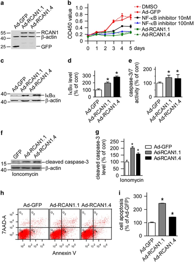
Figure 4.
Overexpression of RCAN1 decreased lymphoma Raji cell viability (a) Expression of RCAN1 isoform 1 and 4 in Raji cells by RCAN1.1 and RCAN1.4 adenovirus. Anti-GFP antibody was used to detect the expression of RCAN1. β-actin was used as loading controls. (b) RCAN1 reduced Raji cell viability. Raji cells infected with adenovirus overexpressing RCAN1.10 and 100 nM NF-κB activation inhibitor (Calbiochem, #481406) were used as positive controls. Dimethyl sulfoxide (DMSO) was the control for NF-κB inhibitor. Ad-GFP infected cells was used as negative control for RCAN1. CCK8 cell viability was measured by the absorbance at 450 nm (ref: 650 nm) at indicated times after adenovirus infection. P<0.0001 by two-way ANOVA. (c) Anti-IκBα was used to detect endogenous IκBα protein level in Raji cells infected with RCAN1 adenovirus. β-actin was used as loading controls. (d) Quantification of c. Values represent mean±S.E.M.; n=3; *P<0.001 by Student's t-test. (e) Raji cells were infected with Ad-GFP, Ad-RCAN1.1-GFP and Ad-RCAN1.4-GFP. Caspase-3/7 activity was measured 48 h after infection. Values represent mean±S.E.M.; n=3; *P<0.05, by Student's t-test. (f) Anti-cleaved caspase-3 (Asp175 from CST) was used to detect the cleaved caspase-3 protein level in Raji cells that were infected with RCAN1 adenovirus and treated with 5 μM ionomycin. β-actin was used as loading controls. (g) Quantification of f. Values represent mean±S.E.M.; n=3; *P<0.001 by Student's t-test. (h) Raji cells infected with RCAN1 adenovirus were stained with 7-AAD and Annexin V and analyzed by FACS to detect cell apoptosis. (I) Quantification of h. Values represent mean±S.E.M.; n=3; *P<0.001 by Student's t-test

