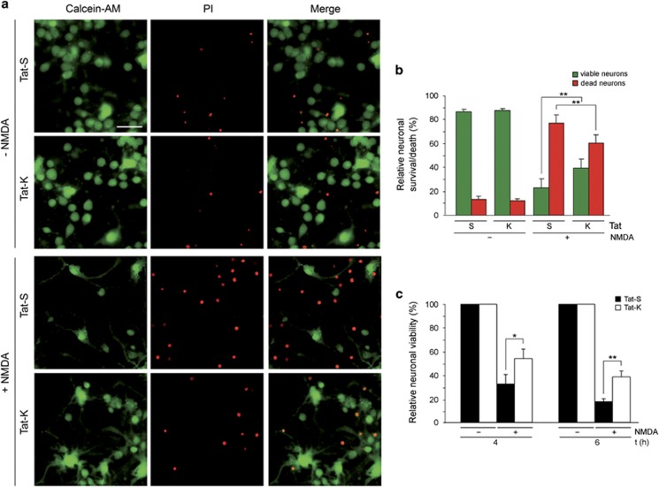
Figure 5.
Neuroprotective effect exerted by Tat-K transduction on excitotoxic neuronal death. (a) Effect of Tat-K on cell death induced by NMDAR overactivation. Primary cultures of cortical neurons (DIV 14) were pre-incubated for 1 h with Tat-S or Tat-K (25 μM) and treated with NMDA for 4 h or left untreated. Next, viable and dead cells were visualized by simultaneous fluorescence staining with calcein-AM (1 μM; green) and propidium iodide (PI; 0.5 mM; red), respectively, for 15 min at 37 º C in the dark. Representative images for each condition are shown. Scale bar, 25 μm. (b) Quantitation of Tat-K effect on the relative percentage of viable/dead neurons found in excitotoxic conditions. A minimum of five random fields containing at least 500 neurons each were counted per experimental condition. The percentage of viable (green bars) versus death (red bars) neurons was expressed relative to the total number of neurons examined, arbitrarily given a 100% value. Average of seven independent experiments with S.E.M. is represented. Statistical significance was determined by paired Student's t-test (**P<0.01). (c) Quantitation of the effect of Tat-K on neuronal viability after induction of excitotoxicity. Cultures of cortical neurons were pre-incubated for 1 h with Tat-S or Tat-K (25 μM) and treated with NMDA for 4 or 6 h as before or left untreated. Neuronal viability was established by MTT reduction assay after subtracting the contribution of the glial cells present in the mixed cultures to absorbance as indicated in the Methods section. For each time point, viability of neurons treated with NMDA in the presence of Tat-S or Tat-K is represented relative to that of neurons incubated with the same peptide but no NMDA, arbitrarily given a 100% value. Average of five independent experiments with S.E.M. is given. Statistical significance was determined by paired Student's t-test (*P<0.05, **P<0.01)

