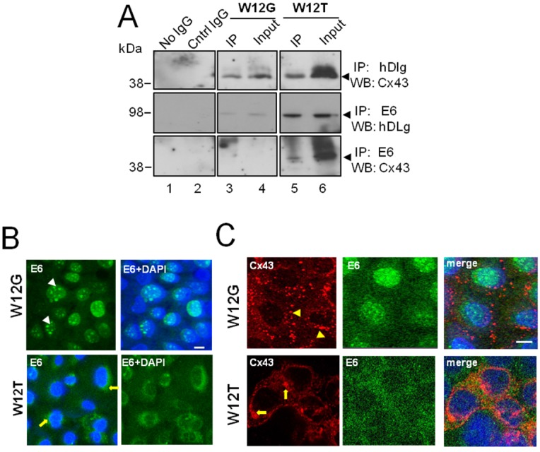
Figure 2.
HPV16 E6 associates with Cx43 in W12T cervical cancer cells. (A) Co-immunoprecipitation of E6 with hDlg and Cx43 in W12G non-tumour and W12T tumour cells. Antibodies used in immunoprecipitation (IP) and the antibodies used to probe western blots (WB) are indicated on the right hand side. No IgG, beads alone used in the immunoprecipitation. Cntr IgG, a matched antibody isotype negative control immunoprecipitation. Input, 10% of the volume of cellular extract as used in the co-immunoprecipitation experiments; (B) Confocal immunofluorescence microscopy imaging showing E6 (green) in the nucleus of W12G cells (arrowheads) but in the cytoplasm of W12T cells (arrows); (C) Confocal immunofluorescence microscopy imaging of Cx43 (red) in W12G cells located in large punctuate gap junction plaques (arrowheads) on the membrane. In W12T cells membrane Cx43 (red) is reduced and some perinuclear staining is detected (arrows). E6 is shown in green. Nuclei are stained with DAPI (blue). Bar = 10 µM.

