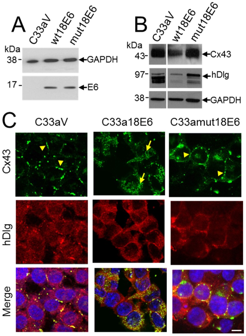
Figure 5.
C33a cells expressing a mutant HPV18 E6 that cannot bind hDlg display membrane Cx43. (A) Western blot analysis of FLAG E6 expression in C33a cells stably transfected with vector alone (C33aV), vector expressing HPV18E6 (wt18E6) or a mutant HPV18 E6 that does not bind hDlg (mut18E6); (B) Western blot analysis of levels of Cx43 and hDlg in C33aV, C33a wt18E6 and C33a mut18E6. GAPDH is used as a loading control for the blots in (A,B); (C) Confocal immunofluorescence microscopy imaging of Cx43 (green) and hDlg (red) in C33aV cells, C33a18E6 cells and C33amut18E6 cells (cells expressing C-terminal mutated 18E6 that cannot bind hDlg). Membrane gap junction plaques are indicated with arrowheads. The arrow in B indicates cytoplasmic Cx43 staining. Nuclei are stained with DAPI. Bar = 10 µM.

