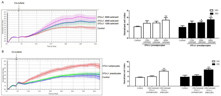
Figure 6.
Real-time monitoring of human umbilical vein endothelial cells (HUVECs) cultured with 3T3-L1 cells. HUVECs were seeded into E-plates at equal densities of 6000 cells/well. (A) 3T3-L1 preadipocytes seeded at different densities into the insert; (B) 3T3-L1 preadipocytes and 3T3-L1 adipocytes seeded into E-plates at equal densities of 1250 cells/well. There were no cells in the insert of control group. After adherence, the insert was added into the E-plates containing HUVECs. Changes in cell index were normalized to the time of co-culture. Experiments were performed in triplicates. The error bars represent the standard deviation (SD) (n = 3); * p < 0.05, ** p < 0.01 vs. the control.

