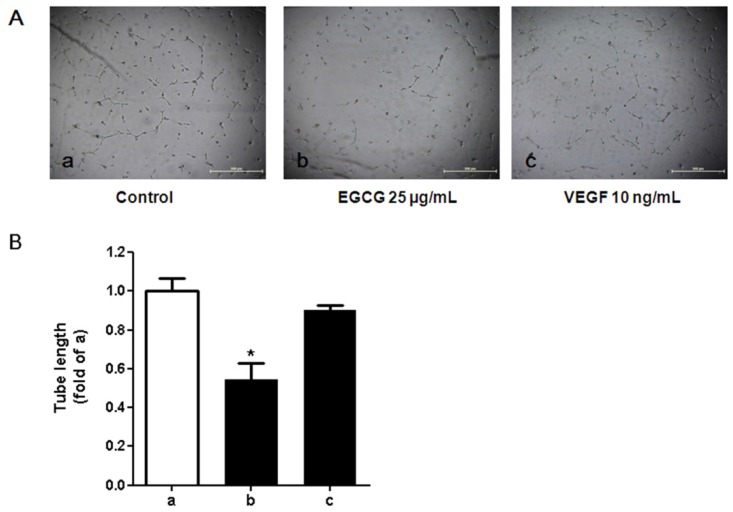
Figure 7.
Effects of the conditioned media cultured with 3T3-L1 adipocytes on tube formation activity in human umbilical vein endothelial cells (HUVECs). (A) Light microscopic images of HUVEC tube formation in various conditions. HUVECs were seeded on Growth Factor Reduced BD Matrigel matrix (BD Biosciences, San Jose, CA, USA) with conditioned medium derived from 3T3-L1 adipocytes in the presence or absence of (−)-Epigallocatechin-3-gallate (EGCG) or recombinant vascular endothelial growth factor (VEGF) protein. After 24 h of stimulation, phase-contrast microscopic low-power fields (×40) were photographed. a: with conditioned media from 3T3-L1 adipocytes in the absence of EGCG; b: with conditioned media from 3T3-L1 adipocytes in the presence of EGCG (25 µg/mL); c: stimulated with recombinant VEGF (10 ng/mL); (B) Tube length of HUVECs stimulated with conditioned medium prepared from 3T3-L1 adipocytes. Total length of capillary tubes formed by HUVECs in 3 different photographs per well was measured using a scale ruler. Bar a: with conditioned media from 3T3-L1 adipocytes in absence of EGCG; bar b: with the conditioned media from 3T3-L1 adipocytes in presence of EGCG (25 µg/mL); bar c: stimulated with recombinant VEGF (10 ng/mL). The error bars represent the standard deviation (SD) (n = 3). * p < 0.05 vs. the control (a).

