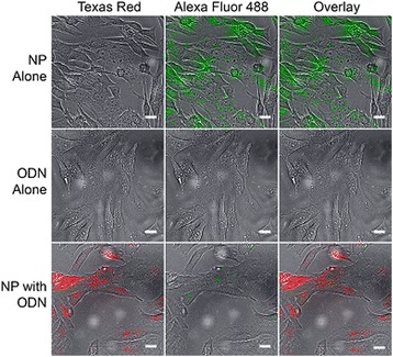
Fig. 6.

Cellular uptake visualization of Alexa Fluor 488 tagged PSA-TMC coated with Texas Red labeled decoy ODNs in SW982 synovial sarcoma cells. Cells were incubated with tagged particles for 45 minutes at 37 °C, and then imaged using an inverted fluorescent microscope. When administered alone, decoy ODN do not register a large signal. However, when delivered using the PSA-TMC carrier system, both red and green signals are clearly seen. NP PSA-TMC nanoparticles, ODN oligonucleotide
