Fig. 1.
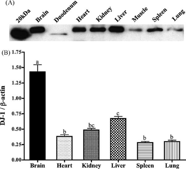
DJ-1 protein is widely distributed in the mouse. (A) A representative blot depicts relative expression of DJ-1 in 25 μg of protein from each respective tissue. (B) DJ-1 protein levels were assayed and are expressed relative to β-actin in each tissue (n ≥ 4), and demonstrate high expression in brain tissue. Bars with differing letters are significantly different from one another (p < 0.05).

