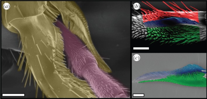
Figure 1.

Antenna cleaner of C. rufifemur ants. (a) Scanning electron micrograph of the antenna clamped by the cleaner (view from the outer=posterior side). (b) Tarsal notch. (c) Tibial spur. Images in (b,c) are coloured to show the bristles (red), the comb (blue) and the brush (green). Scale bars, (a) 150 μm, (b) 100 μm and (c) 50 μm.
