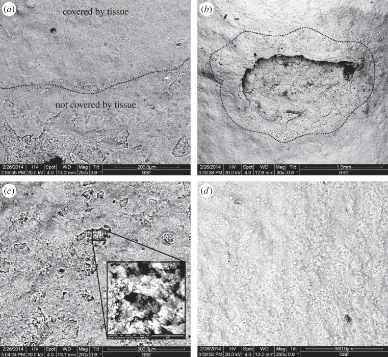
Figure 5.
Back scattered electron emission of Lophelia pertusa skeleton fragments maintained in ΩAragonite < 1. (a) The interface (dashed line) between tissue-protected skeleton (top) and exposed skeleton (bottom). (b) A site of tissue damage on L. pertusa, and subsequent dissolution of skeleton in an otherwise protected area. (c,d) Exposed and tissue-protected sections of skeleton, respectively, with close-up inset.

