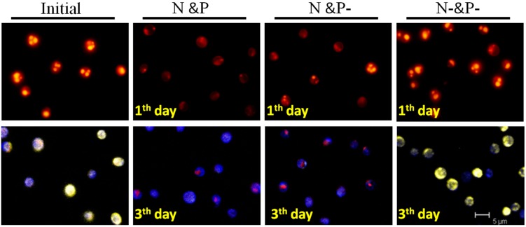
Figure 4.
Observation of neutral lipid and Poly-P using Nile Red and DAPI staining. Above: The golden yellow fluorescence in the images indicates the presence of neutral lipid, and the red fluorescence is the autofluorescence of chlorophyll. Below: Blue fluorescence comes from nucleus staining, bright yellow fluorescence shows the Poly-P, and red fluorescence comes from chlorophyll.

