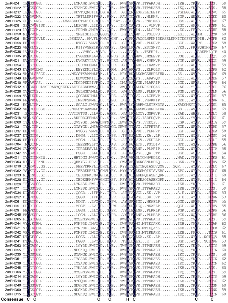
Figure 2.
Multiple sequence alignment of PHD-finger domain of 67 maize PHD proteins. The shading of the alignment presents identical residues in black and similar residues in red, and the high conserved amino are marked at the bottom. The PHD-finger domain of each ZmPHD members is corresponding to the first PHD motif in the upstream of each PHD protein in Figure 3, respectively.

