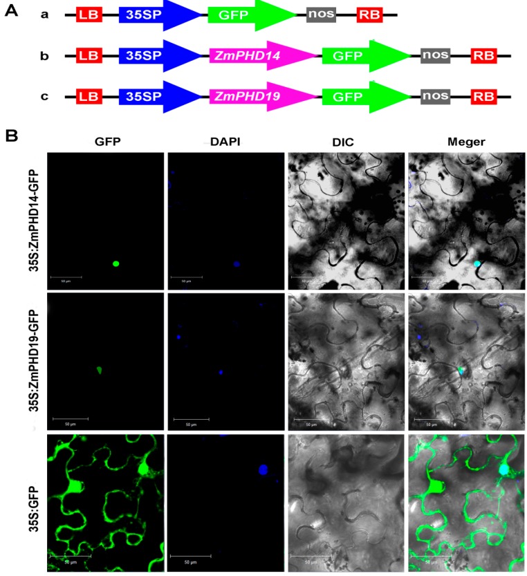
Figure 9.
Subcellular localization of ZmPHD14-GFP and ZmPHD19-GFP fusion protein. (A) Schematic representation of the 35S:GFP, 35S:ZmPHD14-GFP and 35S:ZmPHD19-GFP fusion constructs used for transient expression; and (B) Fusion proteins were transiently expressed under control of the CaMV35S promoter in tobacco leaves and observed under a laser scanning confocal microscope. Green color is GFP protein signal, and blue color represents DAPI stained for nucleus. Bars = 50 μm.

