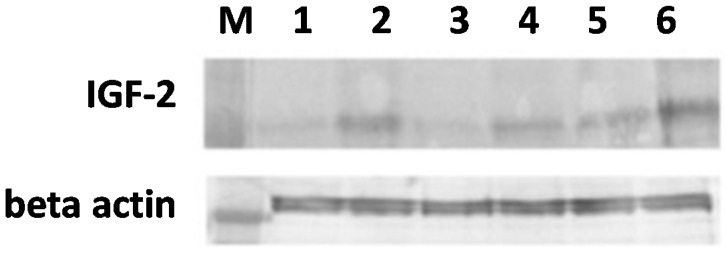
Figure 4.

Western blot analysis of IGF-2 protein in the SW620 human colon adenocarcinoma cell line. Cells were exposed to 3 mM 5-ALA or met-ALA and additionally treated with light (4.5 J/cm2) or not irradiated. Differences between samples were found: Lane 1 shows the band from control cells without treatment; Lane 2 from cells treated with light only; Lanes 3 and 5 from cells incubated with 5-ALA or met-ALA, respectively; Lanes 4 and 6 from cells treated with PDT at the above doses. An increase in IGF-2 was also observed in cells treated with light only (Lane 2) and photosensitizer precursors without light irradiation (Lanes 3 and 5). M, Marker mass.
