Figure 2.
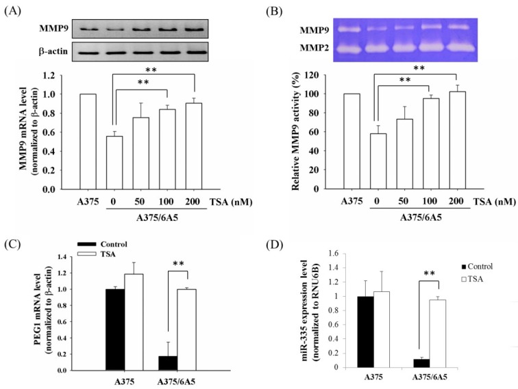
Elevated HDAC activity resulted in the suppressed expression of MMP9 and PEG1 in the PDT-derived variants. (A) The mRNA expression of MMP9 was determined in the A375 and A375/6A5 cells 24 h after treated with different concentrations of TSA. Upper panel is a represented result of the mRNA amount; Lower panel is the average results obtained from three independent experiments (mean ± standard deviation, SD). β-actin served as loading control; (B) gelatinase activity of MMP9 was determined in the A375 and A375/6A5 cells 24 h after various TSA treatments. The upper panel shows a representative gelatin zymography result; The lower panel shows the average of three independent experiments (mean ± SD); (C) mRNA expression of PEG1 was analyzed in the A375 and A375/6A5 cells 24 h after treated with 100 nM TSA. β-actin served as loading control; and (D) the expression of miR-335 was evaluated in the A375 and A375/6A5 cells 24 h after the treatment with 100 nM TSA. Samples were normalized to the small-nucleolar RNA RNU6B. **, p < 0.01.

